A Diagnostic Bull’s-eye? Researchers Use Platelets and Citrate to Take Aim at Lyme Disease
By Michael Lim
1 December 2020
The world may be caught in the grasp of one of the most deadly pandemics in recent history, but COVID-19 is just one zoonotic disease that threatens public health. Industrialization and a warming climate have increased the intermingling of animal and human populations, and with it the risk of disease transmission. This is especially true in the case of Lyme disease, which has increased dramatically in recent years. Now, a study by Dr. Melanie Wills, Scientific Director of the G. Magnotta Lyme Disease Research Lab in the Department of Molecular and Cellular Biology, has developed a new diagnostic method that may improve our ability to detect this complex and potentially devastating infection.
First identified in 1976 in Lyme, Connecticut, Lyme disease is caused by the transmission of bacteria from the genus Borrelia, typically via tick bites. The Centre for Disease Control estimates ~300,000 individuals are diagnosed each year in the US alone. Infections are usually associated with a distinctive “bull’s-eye” pattern rash, but infected individuals may also have no rash at all, or display a mix of different symptoms including headaches, fever, or joint pain.
Diagnostic tests are available, but are far from perfect. They typically involve taking blood samples from patients and testing for antibodies produced in response to Borrelia infection. Unfortunately, the sensitivity of these tests is limited by several factors, including the level of immune response in an individual, the stage of the infection, and the specific strain of Borrelia.
Combined with inconsistent symptoms, this makes early detection difficult – and early diagnosis is critical to successful treatment with antibiotics. Without early intervention, patients can develop serious long-term issues including arthritis, nerve pain, facial palsy, and an irregular heartbeat.
“When I first tell individuals about the state of Lyme disease testing, they often ask ‘how are we still at this stage?’” says Wills. “By the 1990s, there was a sense in some circles that the case was closed on Lyme disease research. Researchers thought, ‘we know the bacteria, we have the antibiotics to treat it. What’s the problem?’ Since then, we have learned just how broad and long-lasting the effects of Lyme disease can be, and how many cases are potentially mis- or undiagnosed.”
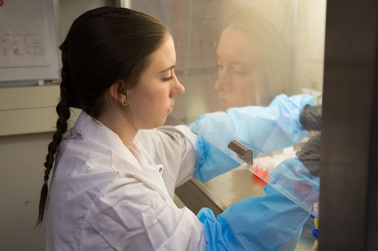
To help spearhead the development of a better diagnostic test, Wills recruited MSc student Victoria Sanderson. In a twist of fate, Lyme disease had a personal connection for Sanderson, as someone close to her had recently been diagnosed. After hearing about the research being done by Wills, she was eager to join the project.
Faced with a wide variety of testing variables to control, Sanderson and Wills decided to focus on two main factors that could be easily adopted by testing clinics around the world: what container to collect sampled blood in, and what portion of blood to test.
Blood sample tubes are typically coated with an anticoagulant, but this can inhibit Borrelia growth, reducing the amount of pathogen counted, and thus underestimate the severity of infection. To identify the best type of tube to use in Lyme disease testing, Sanderson examined tubes coated with either an EDTA or citrate anticoagulant, or left uncoated.
After collecting blood from healthy subjects, Sanderson added Borrelia and incubated the samples for several weeks. She then compared the amount of Borrelia in each type of tube.
Although EDTA has clearly been the anticoagulant of choice in Lyme studies, Sanderson found a significant reduction in Borrelia growth after just 7 days compared to both the uncoated tubes and those coated with citrate! In other words, citrate did a better job of allowing normal Borrelia growth, making it more likely to be detected during sample analysis.
Sanderson then turned her focus to the blood samples themselves. She used high speed centrifugation to separate infected blood into three different fractions: whole blood cells, plasma and platelets. Testing the different blood fractions revealed that the platelets had the highest amount of detectable Borrelia, with nearly 10 times the amount of the next highest fraction. This means that focusing testing on the platelet fraction, where the bacteria appear to naturally concentrate, may lead to fewer missed diagnoses.
While additional research is needed to validate this new testing methodology for clinical use, the results offer new hope that a much-needed improvement in Lyme disease diagnostics may be on the horizon.
“We were really just trying to question the status quo,” says Sanderson. “To take a step back, and make sure that these diagnostic tests were based on a solid foundation. We may not have solved the problem just yet, but science is incremental. We’re making progress.”
Other major collaborators for this work include I.L. Mainprize, L. Verzijlenberg, R. Walchyshyn, and C.M. Kkursigara from the Department of Molecular and Cellular Biology, and A. Tucker and P. Sathasivam from the Human Nutraceutical Research Unit. Funding for this work was provided by the G. Magnotta Foundation and the Natural Sciences and Engineering Research Council of Canada.
Read the full article in the Journal of Biology.
Read about other CBS Research Highlights.