Study Discovers Unexpected Ability in Cell-trafficking Protein
By Daniel Cervone
February 23, 2018

A novel cell-trafficking protein may have an unexpected ability to change how certain molecules are transported in and around cells – a finding that could have implications for diseases like cancer, say researchers in the Department of Molecular and Cellular Biology.
Coordinating the way things are commissioned to move around a cell can be tricky business. Despite lacking the defined infrastructure and painted lines that you may find on city streets, the machinery in a human cell is very efficient at getting things where they need to be, particularly when prompted to do so. A family of proteins known as “Shc” proteins play an important role in this process by coupling external stimuli to internal cellular processes.
Now, a team of researchers led by Prof. Nina Jones has shown that a specific Shc protein, “ShcD”, behaves differently compared to its family members. In a study published in the prestigious Journal of Cell Science, Jones and graduate students Melanie Wills and Hayley Lau characterized this new protein and convincingly demonstrated its ability to change cellular trafficking. Namely, the movement of a molecule called epidermal growth factor (EGF).
EGF is a protein that promotes cell growth. EGF acts like a “key” that binds to a specific receptor or “lock”, EGFR, located on the cell membrane. Once the key fits the lock, this relays a message to other proteins which carry out a very specific effect. The cell can also internalize this “lock and key” complex which either leads to a host of diverse cellular events or gets targeted for death. However, the key can only fit the lock if the lock is available.
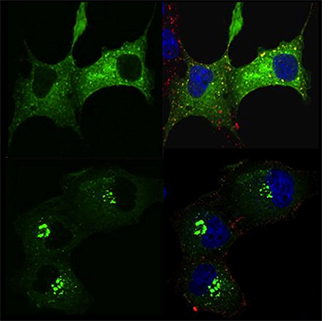
Jones and her colleagues used fluorescently-tagged proteins to track how things moved around both healthy and cancer cell lines and subsequently became active in the presence of different Shc proteins. They were astonished to discover that the ShcD protein restrained the EGFR to the center (nucleus) of the cell – sheltered away from where it needed to be to meet with the key at the cell’s surface.
“EGFR may have sites that ShcD can regulate, somewhat negating the necessity of a ligand (the key) in its entirety – this intrigues us!” says Lau.
The team also found that in ShcD’s presence, the EGFR “lock” tends to be tagged with a molecule called ubiquitin, which is classically considered a signal for its degradation. Lastly, when they modified ShcD’s structure so that it more closely resembled other members of the Shc family, it restored ShcD’s ability to bring EGFR to the cell surface and bring the key into the cell.
Many human cancers (eg. melanoma, glioma) display higher levels of ShcD and researchers aren’t yet sure why.
“ShcD’s properties are very unique to this family of proteins” adds Lau. “We’re excited to move forward in characterizing it and applying what we learn to a disease model”.
The above work was supported by grants from both the Natural Science and Engineering Research Council of Canada (NSERC) and the Brain Tumour Foundation of Canada. Jones is a Tier Two Canada Research Chair (CRC) at the University of Guelph.
Read the full article in the Journal of Cell Science.
Read about other CBS Research Highlights.